Renal Quiz 1
Renal Quiz 1
Uploaded on Nvember 28th, 2022
Dr. Afroza Akhter
Assistant Professor
MBBS, FANMB, Fellow SNUH, S. Korea
Institute of Nuclear Medicine & Allied Sciences (INMAS) Dhaka
Bangladesh Atomic Energy Commission, Bangladesh.
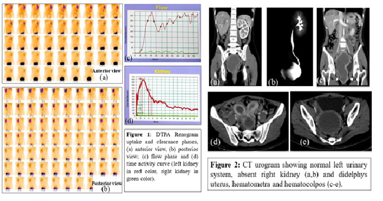
A 14-year-old girl was referred to nuclear medicine department to perform DTPA renogram for non-visualized right kidney with lower urinary tract symptoms and low back pain. DTPA renogram revealed normal functioning left kidney, non-visualized right kidney and a fairly big rim of radiotracer activity in pelvic cavity in early second phase of renogram which was gradually cleared and later obscured by appearance of urinary bladder activity (figure 1). DMSA scan confirmed absent right kidney. Subsequent ultrasonogram and CT urogram (figure 2) revealed single left kidney, absent right kidney and didelphys uterus having hematometra in right moiety and huge hematocolpos causing upward expansion of vaginal cavity up to 5th lumbar vertebra causing her low back pain. Transvaginal surgical exploration revealed obstructed hemi-vaginal septum.
What is the diagnosis?
- Wunderlich syndrome
- Herlyn-Werner-Wunderlich (HWW) syndrome
- Persistent Mullerian duct syndrome (PMDS)
- Mayer-Rokitansky-Kuster-Hauser (MRKH) syndrome
Scroll down for answer
Correct answer-
- Herlyn-Werner-Wunderlich (HWW) syndrome
Herlyn-Werner-Wunderlich (HWW) syndrome is a rare congenital disorder of female urogenital tract characterized by the triad of mullerian duct anomalies including didelphys uterus, obstructed hemivagina and ipsilateral renal agenesis (acronym, OHVIRA syndrome). They usually present with acute or chronic pelvic pain. Diagnosis can be delayed and even misdiagnosed as they have regular menstrual cycle due to obstructed hemivagina. The most common presentation is an abdominal mass secondary to hematocolpos, pain and dysmenorrhea. Ultrasound, MRI & CT scan are the most widely used diagnostic tools. The presenting case didn’t notice for her occasional dysmenorrhea and she had regular menstrual cycle since her menarche at the age of 12. Moreover, hematocolpos was misdiagnosed as right adnexal cystic mass in her previous local ultrasound. She was presented with urinary symptoms and low back pain due to pelvic compression by huge vaginal collection. Though DTPA renogram is not a diagnostic tool for this syndrome, incidental findings of unusual rim of radiotracer activity in the pelvic cavity over the hypervascular thickened vaginal wall in early phase of dynamic renogram helps to explore the final diagnosis as Herlyn-Werner-Wunderlich syndrome.
Wunderlich syndrome is a rare condition in which spontaneous non-traumatic renal hemorrhage occurs into the subcapsular and perirenal spaces. It may be the first manifestation of a renal angiomyolipoma (AML), or the rupture of a renal artery or intraparenchymal aneurysm.
Persistent Mullerian duct syndrome (PMDS) is defined as the presence of Müllerian derivatives, uterus, and fallopian tubes in otherwise normally masculinized 46, XY subjects. It is a rare form of internal male pseudohermaphroditism.
Mayer-Rokitansky-Kuster-Hauser (MRKH) syndrome, also referred to as Müllerian aplasia, is a congenital disorder characterized by aplasia of the uterus and upper part of the vagina in females with normal secondary sex characteristics and a normal female karyotype (46, XX). Uterus agenesis/aplasia in MRKH syndrome has two phenotypic presentations. Two aplastic uterine buds on the pelvic sidewall derived from the Müllerian ducts (often seen in type I) or complete absence of one or both Müllerian ducts (often seen in type II associated with ipsilateral kidney malformations).
References:
- Piccinini, P.S. and Doski, J. (2015). Herlyn-Werner-Wunderlich syndrome: a case
report. Revista Brasileira de Ginecologia e Obstetrícia, 37(4), pp.192–196.
- Del Vescovo, R., Battisti, S., Di Paola, V., Piccolo, C.L., Cazzato, R.L., Sansoni, I., Grasso,
R.F. and Zobel, B.B. (2012). Herlyn-werner-wunderlich syndrome: MRI findings, radiological
guide (two cases and literature review), and differential diagnosis. BMC Medical Imaging,
12(1).
- Cheng, C., Subedi, J., Zhang, A., Johnson, G., Zhao, X., Xu, D. and Guan, X. (2019).
Vaginoscopic Incision of Oblique Vaginal Septum in Adolescents with OHVIRA
Syndrome. Scientific Reports, [online] 9(1).
- Elmas, N.Z., Esmat, H.A., Osmani, G.M., Ozcan, B. and Kızılay, F. (2020). Female form of
persistent Müllerian duct syndrome: A rare case report and review of literature. International
Journal of Surgery Case Reports, [online] 77, pp.298–302.
- Jha, P. (n.d.). Wunderlich syndrome | Radiology Reference Article | Radiopaedia.org. [online]
Radiopaedia. Available at: https://radiopaedia.org/articles/wunderlich-syndrome.
- Herlin, M.K., Petersen, M.B. and Brännström, M. (2020). Mayer-Rokitansky-Küster-Hauser
(MRKH) syndrome: a comprehensive update. Orphanet Journal of Rare Diseases, 15(1).