Neurology Case 3
Neurology 3
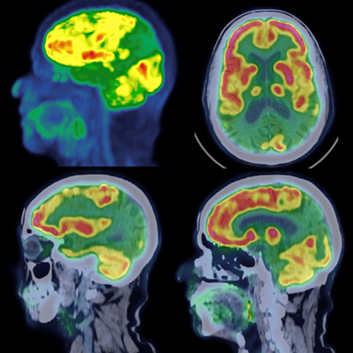
A 69-year-old man with memory loss underwent 18F-FDG-PET/CT brain imaging. Selected images are demonstrated underneath. Based on the findings, what is the most probable cause for the observed appearance of the brain in this examination?
A) Frontotemporal dementia
B) Vascular dementia
C) Alzheimer’s disease
D) Lewy body dementia
Scroll down for answer
Answer: C (Alzheimer’s disease)
Discussion:
Memory loss can arise from a range of underlying conditions, including neurodegenerative diseases. One potential method to detect these disorders earlier is through the18F-FDG PET/CT, which provides valuable insights into metabolic patterns in the brain.
Alzheimer’s disease: This is the most common cause of memory loss in older adults. On 18F-FDG PET/CT, Alzheimer’s disease typically shows hypometabolism (reduced glucose uptake) in the temporoparietal cortex and posterior cingulate gyrus.
Frontotemporal dementia: This type of dementia often presents with behavioral and language changes in addition to memory impairment. 18F-FDG PET/CT may show frontotemporal hypometabolism, involving the frontal and/or temporal lobes.
Vascular dementia: Memory loss due to vascular causes can result from multiple small strokes or chronic cerebral hypoperfusion. PET/CT findings may reveal focal areas of hypometabolism or a more diffuse pattern, depending on the extent and location of vascular damage.
Lewy body dementia: Lewy body dementia is characterized by cognitive decline, visual hallucinations, and parkinsonism. On 18F-FDG PET/CT, it can show a pattern of cortical hypometabolism, particularly in the occipital and parietal lobes.
Other neurodegenerative conditions: There are other less common neurodegenerative diseases, such as Parkinson’s disease, degeneration, and progressive supranuclear palsy, which can also cause memory loss and may exhibit characteristic patterns of hypometabolism on PET/CT.
18F-FDG PET/CT is a highly useful imaging modality for the diagnosis of primary neurodegenerative disorders. It effectively identifies patterns of cerebral glucose metabolism changes that serve as valuable imaging biomarkers, aiding clinicians in the clinical diagnosis of dementia-causing neurodegenerative diseases.
References:
1. Brown RK, Bohnen NI, Wong KK, et al. Brain PET in suspected dementia: patterns of altered FDG metabolism. Radiographics. 2014 May-Jun;34(3):684-701.
2. Shivamurthy VK, Tahari AK, Marcus C, et al. Brain FDG PET and the diagnosis of dementia. AJR Am J Roentgenol. 2015 Jan;204(1):W76-85.
Submitted by:
Dr. Harish Goyal
MD DNB FANMB FEBNM MNAMS
Assistant Professor
Department of Nuclear Medicine
AIIMS Raipur, India