Dr. Harmandeep Singh & Dr. Harpreet Singh
Department of Nuclear Medicine,
PGIMER, Chandigarh
A 54 year old female patient presented with complaints of persistent cough and expectoration since 3 months. CT scan of the chest revealed large mass in the right lung field suggestive of malignancy. Biopsy from the mass was inconclusive.
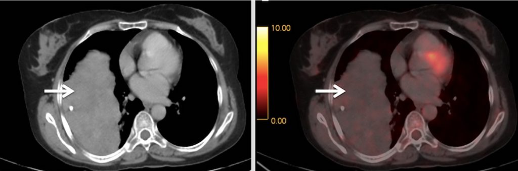
18F-FDG PET/CT was done for lesion characterization.
What is the most probable diagnosis out of the following on basis of 18F-FDG PET-CT findings?
Pulmonary hamartoma
Liposarcoma
Squamous cell cancer
Solitary fibrous tumor
Scroll down for answer
Answer D (Solitary fibrous tumor)
18F-FDG PET-CT images reveal well-defined pleural based lobulated soft tissue mass of size 16×8 cm involving the right lung with low grade FDG avidity (SUVmax 1.6) and punctate calcification within the mass. Repeat biopsy from the mass revealed solitary fibrous tumor.
Discussion:
Pulmonary hamartomas are benign focal lesions which present as a solitary pulmonary nodule (SPN) with a smooth edge with collection of fat or fat alternating with foci of calcification and show no/low grade FDG avidity.
Primary thoracic liposarcomas are very rare and mostly resemble lipomas on CT (areas of lipid attenuation), except for the presence of thick septa and nodular soft tissue elements. Some variants may not show typical features of lipomatous tumors on imaging. Typically, they are low grade neoplasms with low grade FDG avidity. However, Presence of FDG uptake and soft tissue attenuation in thoracic lesions with fatty attenuation indicates malignant liposarcoma.
Squamous Cell Carcinomas (SCC) of lung are predominantly associated with smoking history and present as large cavitatory tumors in the central parts of lung and demonstrate high metabolic activity on PET-CT.
Solitary fibrous tumors (SFT) are rare mesenchymal neoplasms, commonly arising from the pleura and present as well-defined lobulated intrathoracic masses of varied sizes. Larger tumors may show cystic degeneration, calcification and necrosis. On 18F-FDG PET-CT, benign SFTs usually demonstrate low grade tracer avidity while hypermetabolism may be helpful feature in identifying malignant disease. 18F-FDG PET/CT can be used for characterization of thoracic/lung masses.
References:
1. Gaerte S, Meyer C, Winer-Muram H, et al. Fat-containing Lesions of the Chest. RadioGraphics. 2002;22:S61-S78. 2. O’Regan K, Jagannathan J, Krajewski K, et al. Imaging of Liposarcoma: Classification, Patterns of Tumor Recurrence, and Response to Treatment. Am J Roentgenol. 2011;197:37-43. 3. Ambrosini V, Nicolini S, Caroli P et al. PET/CT imaging in different types of lung cancer: An overview. Eur J Radiol. 2012;81:988-1001. 4. Ginat DT, Bokhari A, Bhatt S, et al. Imaging Features of Solitary Fibrous Tumors. Am J Roentgenol. 2011;196:487-495.