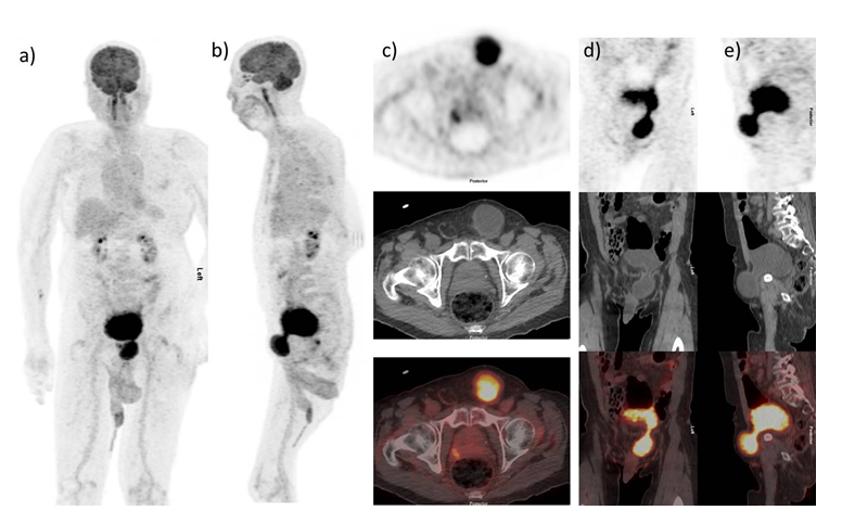
a) FDG activity is related to Inguinal herniation of urinary bladder
a) b) MIP images demonstrate intense uptake in the left inguinal region which is as intense as bladder activity. c) Axial unenhanced CT demonstrates the inferior aspect of the bladder herniating through the left inguinal canal. Axial fused PET-CT images show intense radiotracer uptake in the hernia sac with standard uptake value (SUV) similar to the pelvic bladder activity. The inguinal column of tracer is seen tapering away from the neck, with stagnant activity in the contracted part of the bladder. There is connection of the urinary bladder and hernia sac is best appreciated on sagittal and coronal images (d) and e).
Bladder involvement in inguinal hernia is infrequent, occurring in up to 4% of cases [1 ,2 ]. Most patients are asymptomatic and are diagnosed incidentally on diagnostic imaging [ 3]. They are however at increased risk of acute renal failure, traumatic rupture, fistula formation, bladder calculi, infection and rarely malignancy [4 , 5]. In patients with a narrow hernia neck, voiding can lead to significantly higher activity levels in the herniated bladder relative to the more efficiently emptying native bladder. Interpretation are even more challenging if there is a narrow hernia neck which may result in two separate foci of urinary activity.
Increase FDG uptake is observed in the kidneys, ureters, and bladders because of normal urinary excretion. The differential diagnosis of FDG uptake in the inguinal canal includes testicular cancer, inflammation and urine skin contamination artifact, metastatic lymph nodes, hernia with bowel loops as contents, and inguinal herniation of the urinary bladder. 18 F-FDG-PET-CT is not a primary modality for diagnosing bladder herniation. Few reports have been published previously of incidentally detected herniated urinary bladder on PET-CT [6 ,7 ].
Inguinal herniation of urinary bladder is uncommon and usually an incidental finding in asymptomatic patients. In some of these patients, residual urine volume and consequently, urinary tracer activity can be higher in the herniated bladder then the native bladder, in which case interpretation can be challenging. It is important to important remember that whilst 18F-FDG-PET-CT has a similar spectrum of urinary activity; morphological correlation on hybrid imaging is invaluable in ensuring the physiological nature of uptake.
References:
- Fuerxer F, Brunner P, Cucchi JM, et al. Inguinal herniation of a bladder diverticulum. Clinical Imaging 2006;30:354–6.
- Schaeffer EM, Bhayani SB. Inguinal bladder hernia. Urology. 2003;62: 940.
- Andac N, Baltacioglu F, Tuney D, Cimsit NC, Ekinci G, Biren T. Inguinoscrotal bladder herniation: is CT a useful tool in diagnosis? Clin Imaging 2002;26:347–8.
- Wagner A, Arcand P and Bamberger M. Acute renal failure resulting from huge inguinal bladder hernia, Urology 2004;64:156–7.
- Minordi LM, Mirk P, Canadé A, et al. Massive inguinoscrotal vesical hernia complicated by bladder rupture: preoperative sonographic and CT diagnosis. Am J Roentgenol 2004;183:1091–2.
- Vadi SK, Mittal BR, Singh H, et al. incidental detection of urinary bladder herniation in 18F-fluorodeoxyglucose positron emission tomography/computed tomography mimicking as metastatic deposit in the inguinal canal. Indian J Nucl Med. 2019;34:247–8.
- Usmani S, Marafi F, Ahmed N, et al. Inguinal Herniation of Urinary Bladder on F-18 Sodium Fluoride (NaF) PET-CT. Nucl Med Mol Imaging. 2017;51:368-370.