Oncology Case 2
April 15, 2020
Oncology Case 2
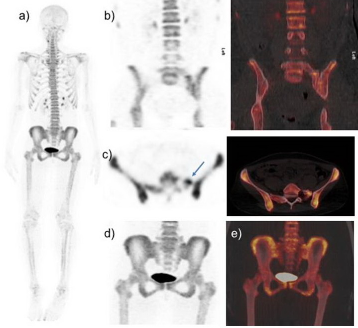
28-years old women has recently diagnosed with breast cancer. She is complaining of backpain. Blood profiles are normal. 18F-NaF-PET-CT performed after injection of 3.27 mCi 18F-NaF for skeletal staging. 18F-NaF-PET-CT demonstrate increase radiotracer uptake is seen at superior aspect of left sacrum. What is the diagnosis ?
A. Skeletal metastasis
B. Osteomyelitis
C. Bertolotti’s syndrome
D. Osteitis condensans ilii
Scroll Down for answer
Answer C; Bertolotti’s syndrome
- 18F-NaF images show broad left transverse process of L5 vertebra forming diarthrodial joint with the sacrum with increase tracer uptake. The focus of increased radiotracer activity corresponds to hemisacralized left transverse process of L5 (Type IIa LSTV). Findings are negative for bone metastasis.
- Osteomyelitis is unlikely due to blood investigations are normal.
- Bertolotti’s syndrome (BS) is a form of low backpain associated with lumbosacral transitional vertebrae (LSTV). LSTV is an anatomical variation of the fifth lumbar vertebra in which an enlarged transverse process can form a joint or fusion with the sacrum or ilium [[i]]. This congenital abnormality occurs in 4%–30% of the general population [[ii]]. The Castellvi system is most commonly used for morphologic classification of LSTV [[iii]]. It defines types I–IV, with type I characterized by an enlarged (>19 mm) transverse process, type II representing vertebra with an incompletely fused broad transverse process forming a diarthrodial joint with the sacrum, and type III having complete fusion of the transverse process with the sacrum. Under each type, unilateral findings are denoted as subtype a, and bilateral findings are denoted b. Type IV consists of a unilateral complete fusion with contralateral incomplete fusion (type III on one side and type II on the other).
Bertolotti’s syndrome is an important cause of low back pain in young patients [[iv]]. The presence of pseudoarticulation has been suggested to contribute to the pain, but there are conflicting reports as well [[v]].
- Osteitis Condensans Ilii (OCI) is a benign cause of axial low back pain [[vi]]. Although no clear etiology has been identified, the prevailing theory is that mechanical strain affects the auricular portion of the ilium and causes premature arthritis [[vii],[viii]]. Characteristic radiographic findings of OCI are well-defined triangular-shaped unilateral or commonly bilateral sclerosis of the lower part of the iliac bone adjacent to a normal sacroiliac joint with normal inflammatory parameters [[ix]]. No characteristic radiological findings are seen in this case.
18F-NaF PET-CT is a sensitive tool for detecting skeletal metastases and has been found to be more sensitive and specific than 99mTc-MDP SPECT in the evaluation of osteoblastic metastases [[x],[xi]]. Hybrid imaging 18F-NaF PET-CT with improves the diagnostic capabilities of by localizing the increased osseous activity more accurately. This case is highlighting the significance of hybrid imaging in the evaluation of low back pain in young breast cancer patient. In young patients with back pain the possibility of Bertolotti’s syndrome should always be taken in account. 18F-NaF PET CT can be useful in evaluating back pain and 18F-NaF may be used as an adjunctive biological maker for assessing LSTV as a potential cause of pain. Recent study by Usmani S et al [[xii]] found that 18F-NaF provide molecular knowledge of biomechanical alterations caused by LSTVs and there is strong linear trend between intensity of 18F-NaF uptake and presence of symptoms.
REFERENCES
[i]. Santavirta S, Tallroth K, Ylinen P, et al. Surgical treatment of Bertolotti’s syndrome. Follow up of 16 patients. Arch Orthop Trauma Surg. 1993;112:82–87.
[ii]. Delport EG, Cucuzzella TR, Kim N, et al. Lumbosacral transitional vertebrae: incidence in a consecutive patient series. Pain Physician 2006; 9:53–56.
[iii]. Konin GP, Walz DM. Lumbosacral transitional vertebrae: classification, imaging findings, and clinical relevance.AJNR American journal of neuroradiology. 2010;31:1778–786.
[iv]. Quinlan JF, Duke D, Eustace S: Bertolotti’s syndrome. A cause of back pain in young people.J Bone Joint Surg Br 2006; 88:1183–1186.
[v]. Almeida DB, Mattei TA, Sória MG, et al.: Transitional lumbosacral vertebrae and low back pain: diagnostic pitfalls and management of Bertolotti’s syndrome. Arq Neuropsiquiatr 2009; 67:268–272.
[vi]. Dihlmann W, Osteitis condensans ilii and sacroiliitis. J Rheumatol. 1991 Sep;18:1430-2.
[vii]. Mitra R. Osteitis condensans ilii. Rheumatol Int. 2010;30:293–296.
[viii]. Cidem M, Capkin E, Karkucak M, Karaca A. Osteitis condensans ilii in differential diagnosis of patients with chronic low back pain: a review of the literature. Mod Rheumatol. 2012; 22:467–469.
[ix]. Numaguchi Y. Osteitis condensans ilii, including its resolution. Radiology. 1971;98:1–8.
[x]. Grant FD, Fahey FH, Packard AB, et al. Skeletal PET with 18F-fluoride: applying new technology to an old tracer. J Nucl Med. 2008;49:68–78
[xi]. Even-Sapir E, Metser U, Mishani E, et al. The detection of bone metastases in patients with high-risk prostate cancer: 99mTc-MDP planar bone scintigraphy, single- and multi-field-of-view SPECT, 18F-fluoride PET, and 18F-fluoride PET/CT. J Nucl Med 2006;47: 287–297.
[xii]. Usmani S, Ahmed N, Marafi F, et al. 18F-sodium fluoride bone PET-CT in symptomatic lumbosacral transitional vertebra. Clin Radiol. 2020 [In Press]. DOI: https://doi.org/10.1016/j.crad.2020.03.023