Osteology case 3
Osteology Case 3
Uploaded on June 30, 2020
Case contributed by:
Dr. Smita Chinmay Kulkarni, DNB (Nucl Med), FEBNM, FANMB, MNAMS
Assistant Professor,
Department of Nuclear Medicine and Molecular Imaging,
Amrita Institute of Medical Sciences,
Kochi, Kerala, India
26 year old male patient with long standing deformity with shortening of right lower limb presents to nuclear medicine department with new onset pain in anterior aspect of right thigh. He had no history of trauma. On examination, there was coxa vara on right side.
Plain radiograph of right hip joint and femur showed ground glass matrix with cystic areas, intact thinned out cortex with expansion of bone. No evidence of fracture.
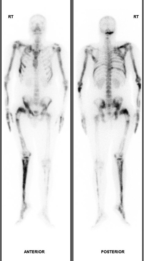
Patient underwent whole body Tc-99m MDP bone scan. Following are the anterior-posterior whole body sweep images.
The patient also underwent an MRI of thigh which showed heterogeneously enhancing intramuscular lesions in right quadriceps muscle consistant with myxoma.
1. What is the diagnosis based on bone scan and clinical features
2. What is the syndrome associated with the disease?
Choices:
Q1.
a. Polyostotic pagets
b. Osteogenesis imperfecta
c. Polyostotic fibrous dysplasia
d. Anterior thigh tumour with skeletal metastases.
Q2.
a. McCune albright syndrome
b. Mazabraud Syndrome
c. Jaffe-Lichtenstein Syndrome
d. Ekman-Lobstein disease.
Scroll down for answer
Answer:
Q1 : c. Polyostotic fibrous dysplasia
Q2 : b. Mazabraud syndrome.
Explanation:
Fibrous dysplasia is relatively uncommon bony disorder where the normal bone is replaced by scar like-fibrous tissue, causing progressive weakening of bone, deformities, limb shortening due to early closure of growth plate and fractures. It can involve single bone (mono-ostotic) or multiple bones (Polyostotic). Classical x-ray feature are presence of ground glass matrix with bony expansion, thinned out cortex. Common sites of involvement include craniofacial bones but sites are not uncommon. When the femur is involved, it produces deformity at the femoral neck causing coxa vara angulation at the hip. This is called Shepherd’s Crooks deformity. The bone scan shows diffuse/ heterogeneous increased tracer uptake in affected bones.
The bone scan findings may overlap with Polyostotic Paget’s disease of bone. However the age of presentation, clinical features and x ray can help to differentiate both entities. In Paget’s disease, x ray shows osteolytic/ (lucent) areas, subsequently appearing as coarsened trabeculae and bony enlargement and sclerotic changes with further progression.
Osteogenesis imperfecta is a genetic disorder which can range from milder form to very severe form, mostly presents very early in life. May be associated with other collagenous pathologies like blue sclera and hearing loss.
Q.2
Mazabraud syndrome is a rare syndrome where Polyostotic fibrous dysplasia is associated with intramuscular mxyoma.
In McCune-Albright syndrome, usually seen in girl child, Polyostotic fibrous dysplasia are associated with cafè-au-lait skin pigmentations, endocrine problems like diabetes, precocious puberty, goitre and firoadenomas of breast.
Osteogenesis imperfecta tarda is also known as Ekman-Lobstein disease.
Jaffe-Lichtenstein Syndrome is a broder term for syndromes associated with fibrous dysplasia.
References:
1. Yevgeniya S. Kushchayeva, Sergiy V. Kushchayev, Tetiana Y. Glushko, Sri Harsha Tella, Oleg M. Teytelboym, Michael T. Collins, Alison M. Boyce. Fibrous dysplasia for radiologists: beyond ground glass bone matrix. (2018) Insights into Imaging. 9 (6): 1035.
2. Zhibin Y, Quanyong L, Libo C, et al. The role of radionuclide bone scintigraphy in fibrous dysplasia of bone. Clin Nucl Med. 2004;29(3):177‐180.
3. https://radiopaedia.org/articles/fibrous-dysplasia.