Answer: C. Camaruti Engelman disease
Discussion:
Camurati-Engelmann disease
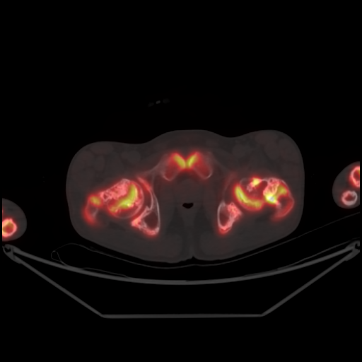
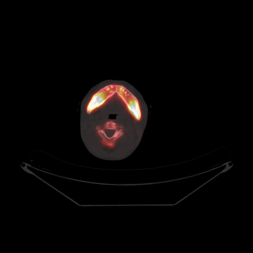
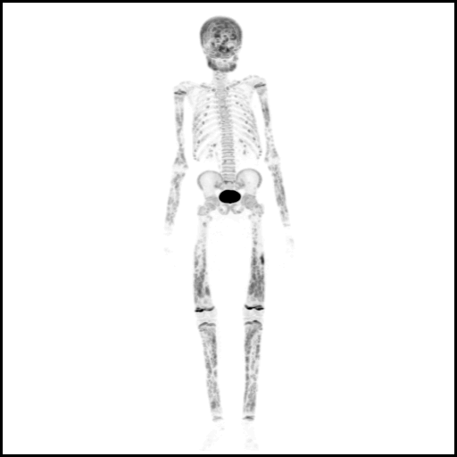
Also called progressive diaphyseal dysplasia, It is a rare sclerosing bone dysplasia resulting from a mutation in TGFB1 gene leading to osteoblastic overactivity. It presents in childhood and common complaints are bony pain and muscle weakness. It worsens with age and can lead to cranial nerve palsies and gait abnormalities. Disease involvement is often bilateral and symmetrical, with predilection for long limb bones. Fusiform bony enlargement with sclerosis in long bones, sparing the epiphysis is seen on X-ray. High tracer uptake in involved bones is seen on 99mTc-MDP scintigraphy or 18F-Fluoride PET.
References:
Weerakkody Y, Sharma R, Bilodeau L, et al. Camurati-Engelmann disease. Reference article, Radiopaedia.org (Accessed on 11 May 2023) https://doi.org/10.53347/rID-10851
Damiá Ade B, Morón CC, Pérez PA et-al. Bone scintigraphy in Engelmann-Camurati disease. Clin Nucl Med. 2010;35 (7): 559-60. doi:10.1097/RLU.0b013e3181e05ea3