Endocrinology 2
May 9, 2020
Case Submitted by:
Dr. Satyawati Deswal
Additional Professor
Dept. of Nuclear Medicine
RMLIMS, Lucknow-226010
India
A 22 years old female presented with generalised body ache for 1 yr and underwent MRI for right upper leg swelling which revealed multiple lobulated exophytic lesions in tibial shaft causing scalloping of adjacent soft tissue and contour bulge and a similar small lesion in proximal shaft of fibula. FNA Cytology showed occasional giant cells along with lymphocytes likely fibrous dysplasia. Serum biochemistries revealed that her serum calcium was 11.30mg/dl (normal 8-10mg/dl), serum PTH was 1453.9 pg/ml (normal 10-55pg/l), serum creatinine was 0.90mg/dl (0.6-1.2mg/dl). A dual phase 99 Tc-sestamibi scanning for parathyroid evaluation was performed.
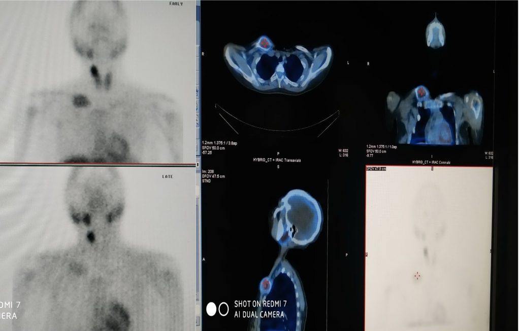
Sestamibi planar imaging at 10 minutes and 120 minutes was done and this showed showed an area of increased uptake at the upper pole of right lobe of thryoid, this persisted at the two hour image while the rest of the thyroid uptake had declined. An abnormal area of increased uptake was seen in the medial end of the rt clavicle. On SPECT-CT the thoracic abnormal area was confirmed to be in the clavicle.
Image on top: left, planar sestamibi 10 minute, anterior projection head, neck and thorax; mid, transverse sestamibi SPECT-CT section through the rt clavicle; right; Coronal sestamibi SPECT-CT.
Images on bottom: left, planar sestamibi 120 minutes anterior projection head, neck and thorax; mid, sagittal section SPECT-CT through the right clavicle; right, SPECT anterior delayed.
What is the diagnosis?
a. Papillary carcinoma thyroid with lymph node metastases
b. Right superior parathyroid adenoma with brown tumor in Medial end of right clavicle.
c. Follicular carcinoma thyroid with skeletal metastases.
d. Double adenoma Parathyroid
Scroll down for answer and discussion.
Correct answer is "B"
Where biochemical screening is a common practice, most patients with primary hyperparathyroidism are asymptomatic when diagnosed. In 1930, Fuller Albright described it as “bones, stones and groans”. Resorption of bone caused by parathormone induced increased osteoclast activity result in lytic and cystic bone lesions known as brown tumors. Parathyroid adenoma is the most common cause of primary hyperparathyroidism. Imaging is done for preoperative localization of adenoma prior to surgery. Tc-99m sestamibi is generally regarded to be the most sensitive and specific imaging modality, especially when it is combined with SPECT CT. For a single parathyroid adenoma, sensitivity is 80% to 100%, with a 5% to 10% false-positive rate (usually with coexistent thyroid nodules). Uptake of 99m Tc-sestamibi has also been reported in brown tumors.
References:
- Primary hyperparathyroidism: Pathophysiology, surgical indication and preoperative work up. Surgery of thyroid and parathyroid glands. Ch 53; pg 486-493
- Radionuclide imaging of parathyroid glands. Seminars in nuclear medicine 35 : 266-276; 2005.
- Primary hyperparathyroidism: Review and recommendation on evaluation , diagnosis and management. A Canadian and international consensus. Osteoporos Int. 2017; 28;pg 1-19.
- Osteitis fibrosa cystica—a forgotten radiological feature of primary hyperparathyroidism. Endocrine. 2017; 58(2): 380–385.
- Clinical implications of brown tumor uptake in whole-body 99mTc-sestamibi scans for primary hyperparathyroidism. Nucl Med Commun. 2011 Aug;32(8):708-15