Osteology Case 9
Dr. Sharjeel Usmani
KBNM, FEBNM,FANMB, CBNC
Consultant Nuclear Medicine
Sultan Qaboos Comprehensive Cancer Care and Research Centre
Muscat, Oman
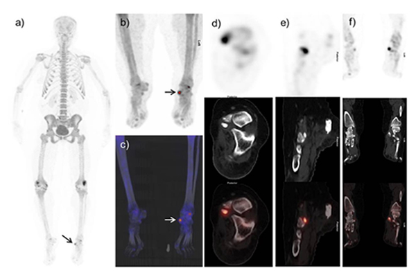
54-years old female with breast cancer with left foot pain had 18F-NaF-PET-CT performed after injection of 6mCi 18F-NaF. Images demonstrate increase radiotracer uptake in the anteromedial aspect of left mid foot region.
What is the diagnosis ?
a) Osteoarthritic/degenerative changes at left talonavicular joint.
b) Fracture at left navicular process.
c) Findings are most probably due to talonavicular coalition; for further radiological evaluation.
d) Findings are likely due to accessory navicular syndrome.
Scroll down for answer
Answer: d
Non-Contrast CT images demonstrate osseous density medial to the left navicular bone which was suggestive of an Type II accessory navicular bone. a-e) Fused NaF-PET-CT shows increased tracer uptake in the articulation with sclerosis between the medial border of the left navicular and an osnaviculare. This would suggest left painful accessory os syndrome.
Os navicular bone is an accessory bone of the foot, which mainly found on the medial side of the proximal navicular bone and in continuity with tibialis posterior tendon. Its incidence is 6-12%. There are three types reported in the literature [1]. Type I is a small sesamoid bone located within posterior tibial tendon. Type II is a triangular or heart-shaped unfused accessory ossification centre, separated from the tuberosity by a 1–2 mm wide synchondrosis. Type III is a prominent tuberosity, thought to be a fused type II accessory navicular bone [2]. Type II and III are mostly symptomatic and associated with clinical manifestations particularly pain. The mechanism of pain in accessory navicular has been attributed to traumatic or degenerative changes at the synchondrosis or to soft-tissue inflammation.
Type II accessory navicular bone may be symptomatic and cause medial foot pain [3]. Plain radiographs may be not helpful in their diagnosis. On the other hand, bone scintigraphy or MR imaging is helpful [4]. MRI findings of painful accessory navicular bone usually include persistent edema pattern in the accessory navicular bone and within the synchondrosis [5]. The indication of three-phase bone scan is to see if the accessory navicular bone is the cause of symptoms [6]. 18F-NaF PET-CT is a sensitive tool for detecting skeletal metastases surpassing conventional bone scintigraphy [7 ]. Encouraging results are also seen in characterizing benign bone pathology [8]. Few studies also highlight the significance of 18F-NaF PET CT in symptomatic accessory bones [9,10 ]. This case is highlighting the importance of hybrid imaging in the evaluation of symptomatic accessory navicular bone.
References:
- Groshar D, Gorenberg M, Ben-Haim S, et al. Lower extremity scintigraphy: The foot and ankle. Semin Nucl Med. 1998;28:62–77
- Miller TT. Painful accessory bones of the foot. Semin Musculoskelet Radiol.2002;6:153–161.
- Mosel LD, Kat E, Voyvodic F. Imaging of the symptomatic type II accessory navicular bone. Australas Radiol. 2004;48:267–271.
- Shah S, Achong DM. The painful accessory navicular bone: scintigraphic and radiographic correlation. Clin Nucl Med. 1999;24:125–126.
- Choi YS, Lee KT, Kang HS, et al. MR imaging findings of painful type II accessory navicular bone: Correlation with surgical and pathologic studies. Korean J Radiol.2004;5:274–9
- Romanowski CA, Barrington NA. The accessory navicular: An important cause of medial foot pain. Clin Radiol. 1992;46:261–4.
- Löfgren J, Mortensen J, Rasmussen SH, et al. A Prospective Study Comparing 99mTc-Hydroxyethylene-Diphosphonate Planar Bone Scintigraphy and Whole-Body SPECT/CT with 18F-Fluoride PET/CT and 18F-Fluoride PET/MRI for Diagnosing Bone Metastases. J Nucl Med. 2017;58:1778-1785.
- Ovadia D, Metser U, Lievshitz G, et al. Back pain in adolescents: assessment with integrated 18F-fluoride positron-emission tomographycomputed tomography. J Pediatr Orthop. 2007;27:90–3.
- Usmani S, Marafi F, Ahmed N, et al. 18F-NaF PET-CT in Symptomatic Fabella Syndrome. Clin Nucl Med. 2017;42:e199-e201.
- Usmani S, Sit C, Gnanasegaran G, et al. Pictorial atlas of symptomatic accessory ossicles by 18F-Sodium Fluoride (NaF) PET-CT. American Journal of Nuclear Medicine and Molecular Imaging. 2017;7:275-282.