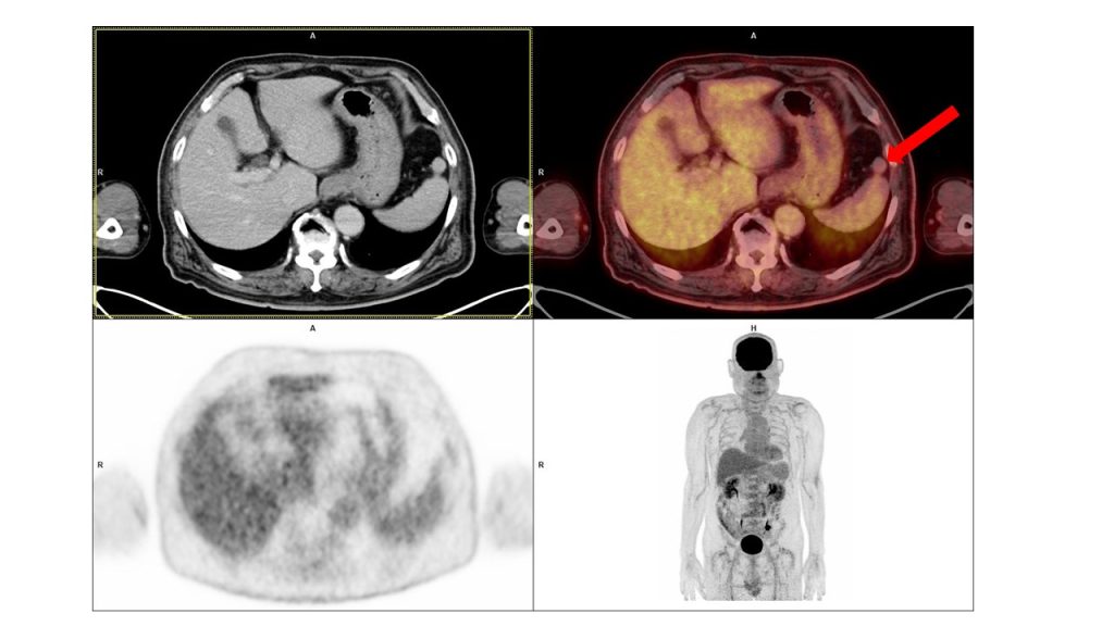
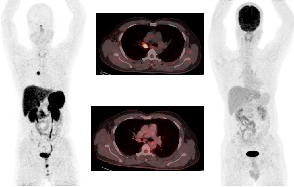
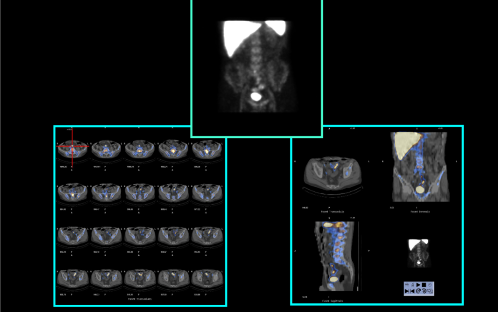
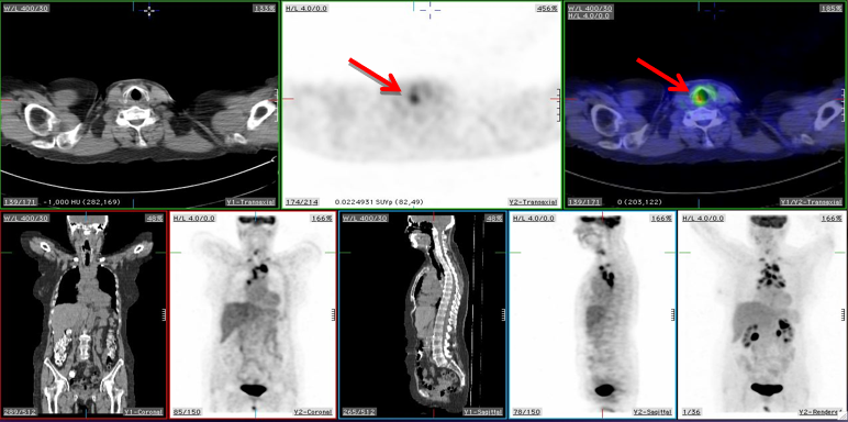
01. What is the anatomical structure/lesion (red arrow) seen on F-18 FDG PET/CT?
A. Peritoneal carcinomatosis B. Splenule C. Splenic cyst D. Nodal metastasis
02. What is the most likely cause of the streaking artifacts seen in the CT images around dense bony structures in this patient?
A. Patient movement during the scan B. Beam hardening artifact C. Inadequate contrast administration D. Detector malfunction
3. What is the fraction of the total radioactivity in the form of the desired radionuclide present in a 23. The dose rate during preparation of a radiopharmaceutical is 1.66 mrem/hr. With this rate reduced by 2 HVLs (half-value-layer), approximately how many hours would it take a worker to accumulate 10 mrems?
A. 3 B. 6 C. 12 D. 24
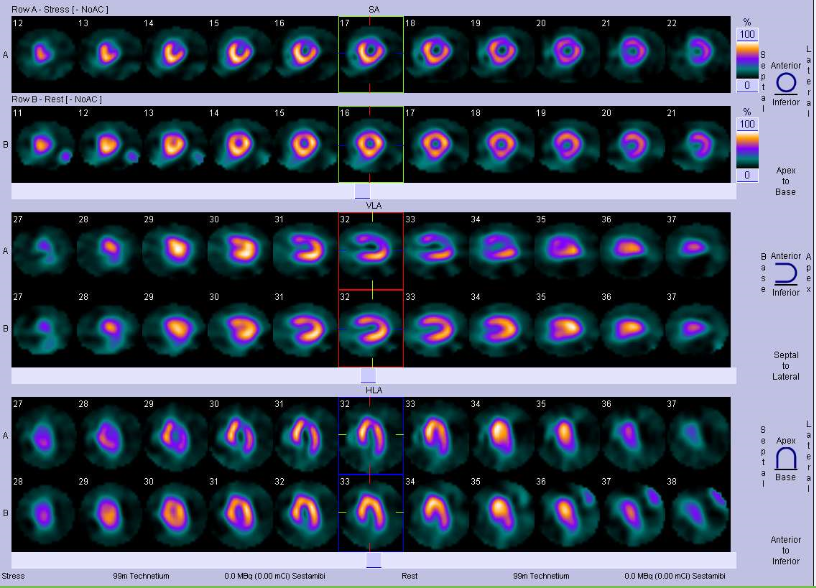
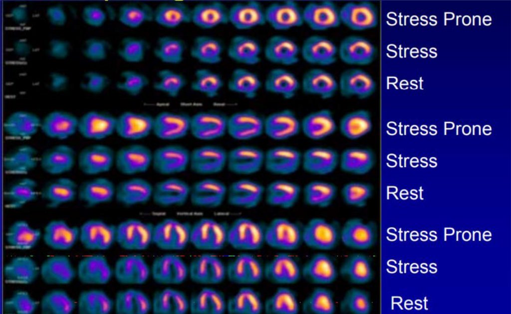
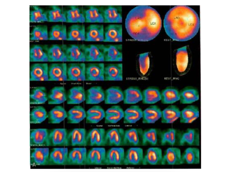
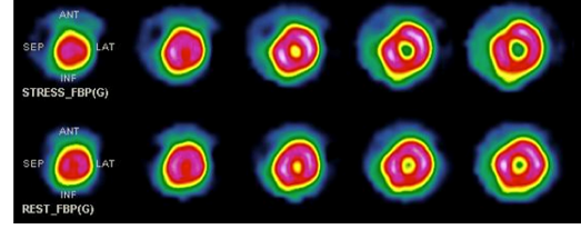
4. A 67-year-old male, with history of hypertension and diabetes mellitus presented with exertional chest pain and dyspnea. His ECG was within normal limits, Echocardiography showed mild LVH with a LVEF of 60%. He was referred for Tc-99m-tetrofosmin stress rest myocardial perfusion imaging. Scan findings are shown below.
The most likely diagnosis is: A. Stress induced ischemia in LAD (Left Anterior Descending) and LCx (Left Circumflex) territories B. Scarred myocardium in LAD and LCx territories C. Stress induced ischemia in RCA (Right Coronary Artery) and LCx territories D. Scarred myocardium in RCA territory
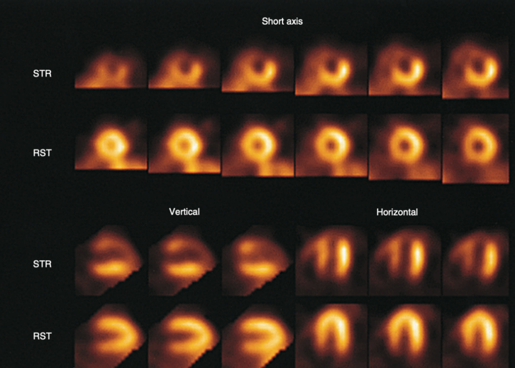
5. A 50-year-old male had an acute coronary syndrome that required hospital care, followed by symptoms of heart failure. Rest ECG showed Q waves in leads II, III, and aVF. Stress-then-rest myocardial perfusion scan using thallium-201 was done, with illustrative scan as follows:
Which of the following statements is true of the case?
A. Angioplasty of the left circumflex artery is indicated B. The extensive defects require coronary artery bypass graft C. Either angioplasty or bypass surgery is appropriate for the right coronary artery D. Optimal medical therapy is appropriate in this case
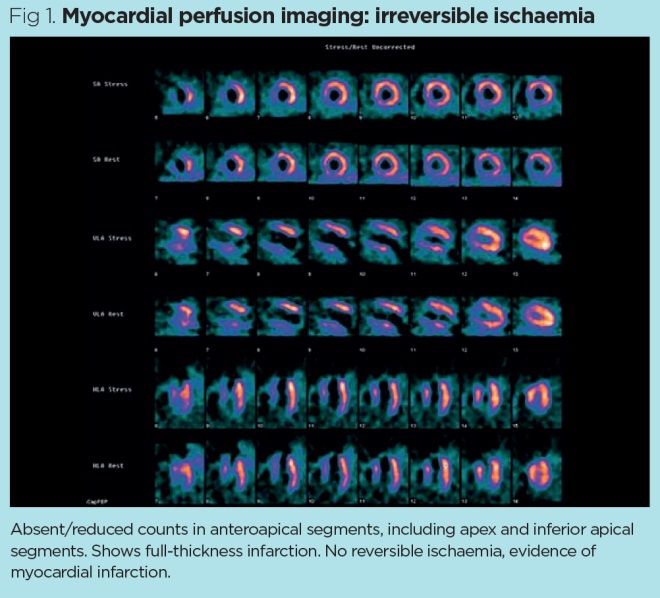
6. A 29-year-old man with atypical chest pain presents for myocardial perfusion imaging. What is the most likely diagnosis?
A. Non-ischaemic dilated cardiomyopathy B. Myocardial infarction in the inferior wall and apex C. No myocardial perfusion abnormality D. Myocardial infarction and peri-infarct ischemia in the RCA territory
7. Which of the following scans shows aTl-201 and exercise?
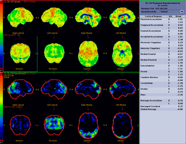
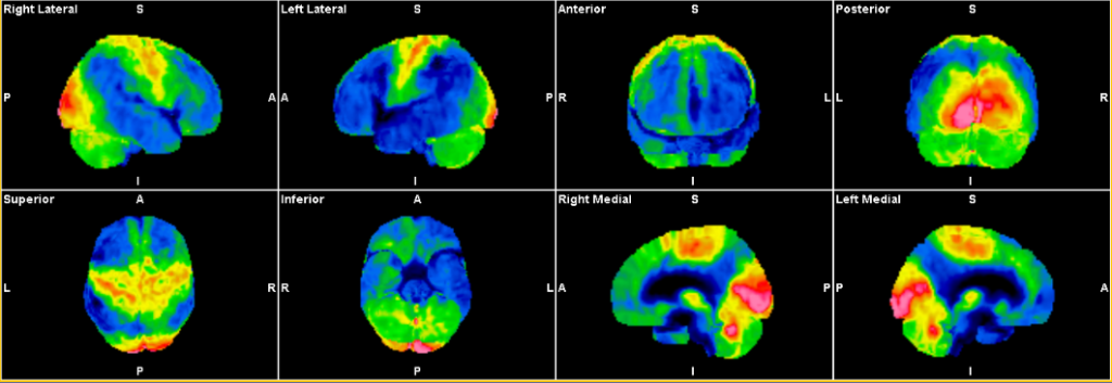
8. A 76-year-old male, presented with history of tremors in the right hand for 1 year and left hand for 6 months. He was treated as Parkinson disease and later he developed memory loss and REM sleep disorder (with positive family history). FDG PET/CT was done and the reconstructed 3D SSP images are illustrated below.
What is the most likely diagnosis? A. Idiopathic Parkinson disease B. Progressive supranuclear palsy C. Diffuse Lewy-body dementia D. Alzheimer’s disease
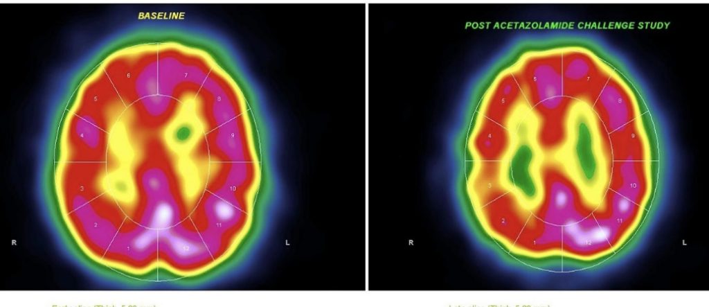
9. A 41-year-old man with right middle cerebral artery stenosis underwent Tc-99m HMPAO brain SPECT imaging at baseline and after acetazolamide challenge. The images are shown below: What is the most likely interpretation of these image findings?
A. Normal cerebrovascular reserve B. Reversible ischemia C. Chronic infarction with no viable tissue D. Impaired cerebrovascular reserve
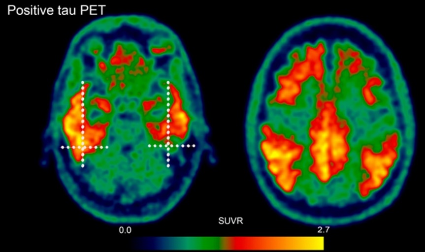
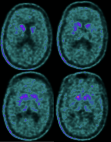
10. A 65-year-old female undergoes F-18 Flortaucipir PET imaging to assess for neurodegenerative disease which is declared as positive. What does a positive scan indicate when using the radiotracer?
A. The presence of amyloid plaques in the cerebral cortex. B. Abnormally increased uptake in the neocortex above the background level, indicative of tau pathology. C. Reduced uptake in the basal ganglia structures such as the caudate and putamen. D. Normal gray-white matter contrasts with no abnormal radiotracer binding.
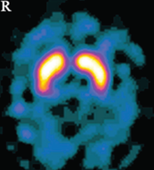
11. Identify the study and its common indication for routine utilization.
A. TRODAT study, for evaluation of pre-synaptic dopaminergic system in evaluation of movement disorders. B. F-18 DOPA study, for evaluation of post synaptic dopaminergic system in evaluation of movement disorders. C. TRODAT study, for evaluation of dementia. D. F-18 Florbetaben study, for evaluation of movement disorders.
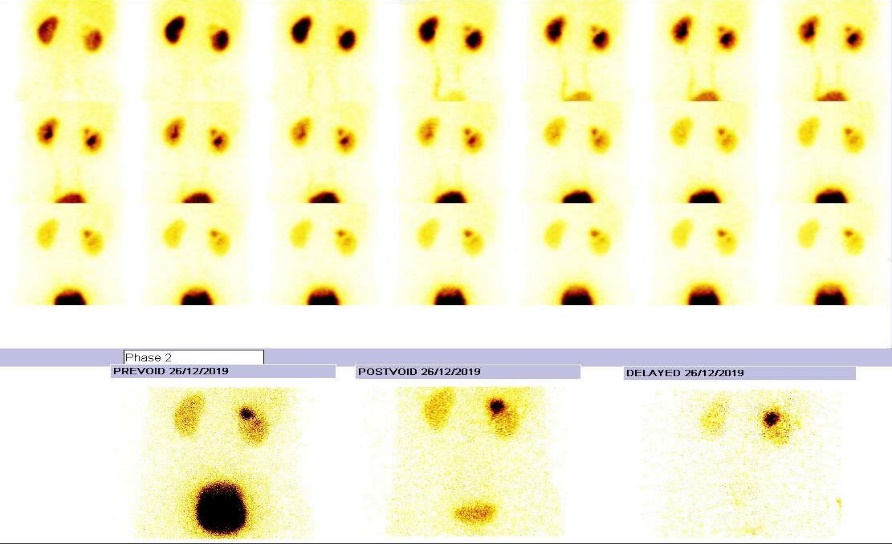
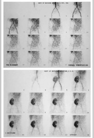
12. A 7-year-old girl underwent a renal dynamic scan due to recurrent urinary tract infections. The posterior view of the scan showed two separate areas of tracer uptake within the right kidney. The images are shown below:
What is the most likely interpretation of these image findings?
A. Horseshoe kidney B. Simple renal cyst C. Duplex collecting system D. Partial Renal agenesis
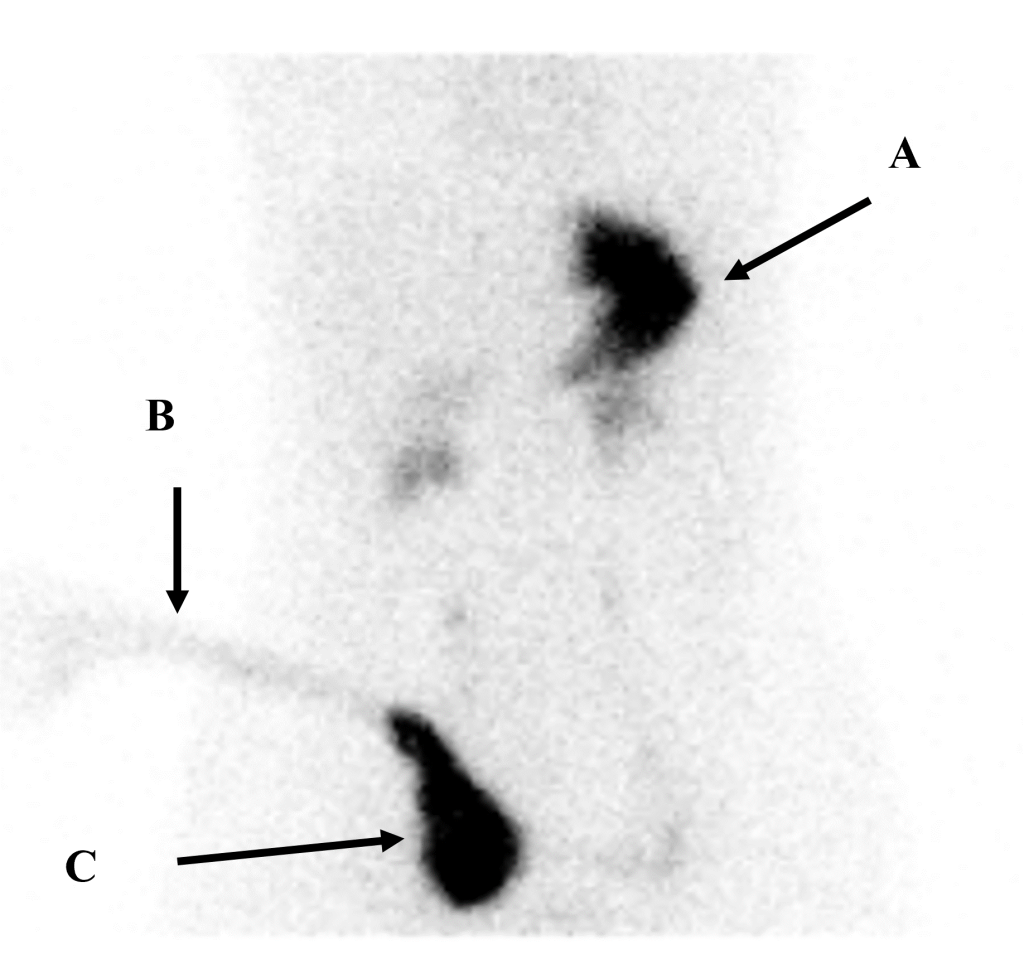
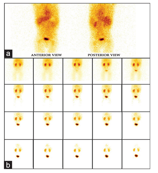
13. The following is the anterior view of Tc-99m pertechnetate abdominal scan in a patient who has undergone bladder augmentation surgery with gastric patch. Select the true statement:
A. B denotes catheter and A denotes left renal activity B. A denotes stomach and C denotes urinary activity in the bladder C. B denotes catheter and C denotes viable gastric patch D. A denotes stomach and C denotes post-surgical collection
14. Which statement about vitamin B12 absorption (Schilling) test is CORRECT:
A. The Schilling test with intrinsic factor can be used to discriminate between intestinal malabsorption and lack of intrinsic factor B. For patients on vitamin B12 injection treatment, it is necessary to wait at least 4 weeks from last injection before performing the test C. Low vitamin B12 absorption when administered with intrinsic factor rules out pernicious anemia D. The test cannot distinguish folic acid deficiency from pernicious anemia
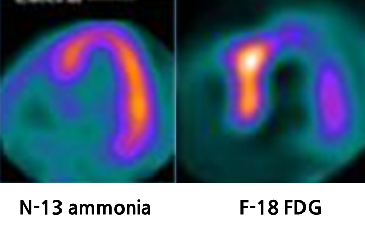
15. A 65-year-old male patient complained of palpitation, syncope, and arrhythmia on ECG. Cardiac PET/CT images are shown below. What is CORRECT statement for the diagnosis ?
A. Diffuse F-18 FDG uptake with preserved perfusion in septum is usually considered as early sarcoidosis. B. Decreased uptake of F-18 FDG with preserved perfusion in lateral wall is usually considered as late sarcoidosis. C. Increaseduptakeof F-18 FDG with a prfusion defect in septum is considered as activesarcoidosis. D. Decreased F-18 FDG uptake and decreased perfusion in apex indicates early sarcoidosis.
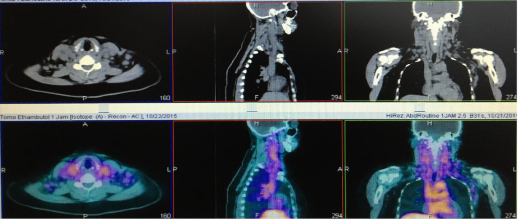
16. A 50-year-old female patient had a history of cervical lymphadenopathy. She underwent Tc-99m ethambutol scintigraphy. What is the CORRECT statement for this patient?
A. Increased uptake in cervical lymph nodes indicate tuberculosis. B. Increased uptake in cervical vertebrae indicate bone metastasis. C. Increased uptake in cervical muscles indicate myositis. D. Increased uptake in cervical lymph nodes indicate lymphoma.
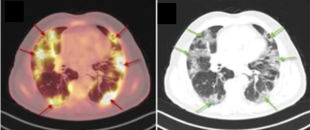
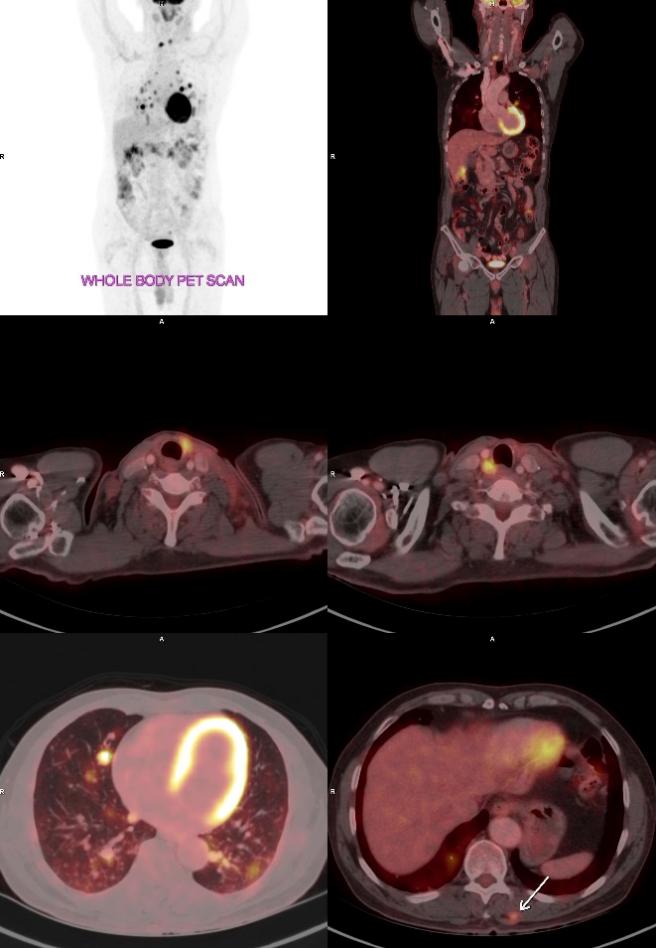
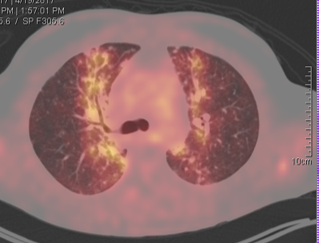
17. A 70-year-old male patient had a history of cough, fever, and chest tightness. What is the diagnosis according to the following F-18 FDG PET/CT images?
A. Sarcoidosis B. COVID-19 infection C. Candida albicans infection D. Pulmonary metastasis
18. A nuclear medicine department schedules patients for various nuclear scans. To evaluate patient satisfaction during their stay at the nuclear medicine department, a systematic random sampling technique is to be used for selecting patient responses on a pre-designed questionnaire. Which of the following approaches best describes systematic random sampling?
A. Randomly select 10 patient records. B. Select every 10th patient record. C. Select the first 10 patient records. D. Select 10 patient records on Mondays.
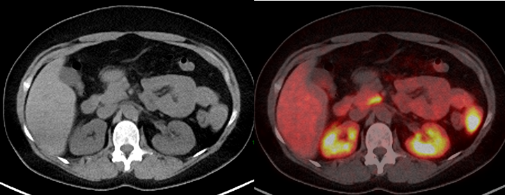
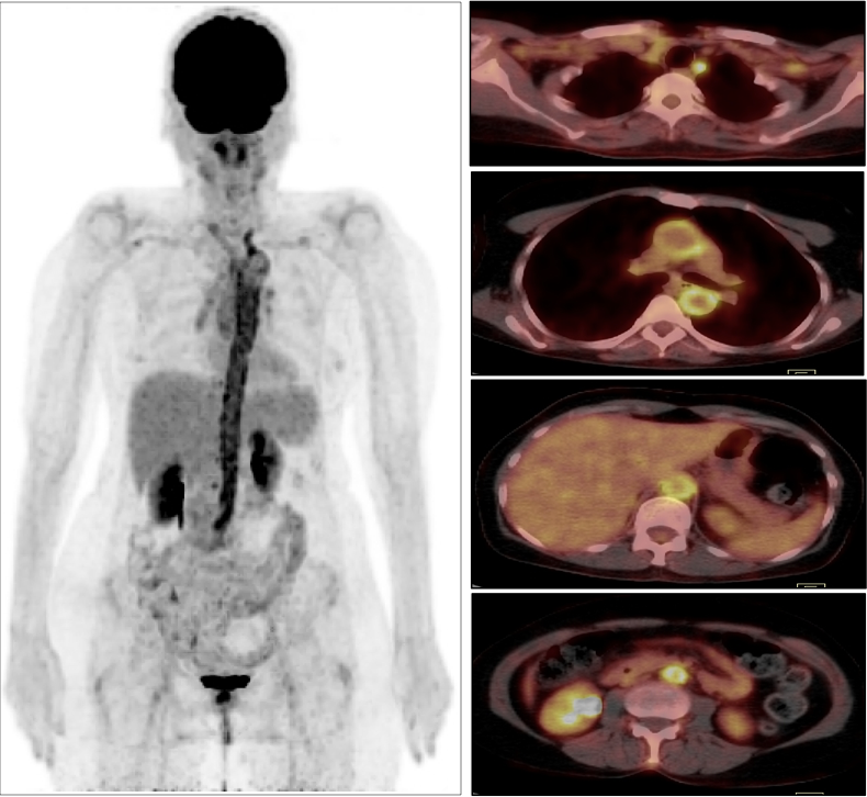
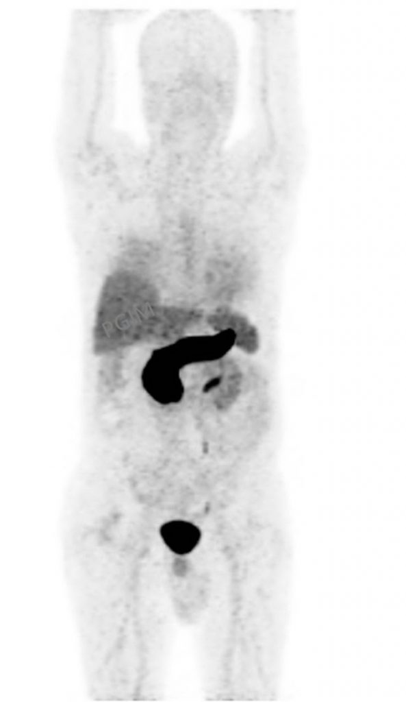
19. A Ga-68 DOTATOC PET/CT scan was performed for re-valuation of post operative medullary thyroid cancer with raised calcitonin level. Focal uptake was seen in the uncinate process of pancreas. The images are shown below:
What is the most correct interpretation of this finding? A. Neuroendocrine tumor of pancreas B. Physiological uptake C. Metastases from medullary thyroid cancer D. Autoimmune disease of the pancreas
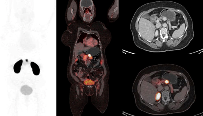
20. A 45-year-old male with a persistent cough and wheezing underwent both Ga-68 DOTANOC PET/CT and F-18 FDG PET/CT scans. The scans showed lesion in the right main bronchus (images shown below).
What is the most likely interpretation of these image findings?
A. Hamartoma B. Sarcoidosis C. Bronchial carcinoid D. Mucus plug
Sample Questions from the 2023 Exam
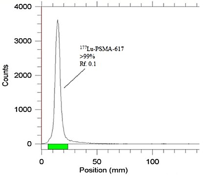
2. Radiopharmacies that prepare Lu-177 PSMA and Lu-177 DOTATATE radionuclide medicines frequently use Instant Thin Layer Chromatography (ITLC) for quality control. What is determined by this test?
A. Radio-nuclide purity of radiopharmaceutical B. Radio-chemical purity of radiopharmaceutical C. Chemical purity of radiopharmaceutical D. Sterility and apyrogenicity of radiopharmaceutical
3. A 55-year-old man with recurrent chest pain had an exercise stress SPECT myocardial perfusion study. The images are shown below:
A. Left anterior descending coronary artery B. Left circumflex coronary artery C. Right coronary artery D. A and C
4. A 67-year-old woman with typical chest pain, dyspnea, and a history of 10-year-old hypertension was referred for MPI. The images are shown below:
Which cardiac artifact is more likely? A. Center of rotation artifact B. Motion artifact C. Attenuation artifact D. Interfering GI activity
5. A 62-year-old male presented with a history of tremors and stiffness in the right upper limb. F-DOPA PET was done. Which of the following is TRUE?
A. Findings are consistent with essential tremors. B. Both Idiopathic Parkinson’s disease and Progressive supranuclear palsy can show these findings. C. Dementia with Lewy bodies and Parkinson’s disease dementia can be differentiated based on these findings. D. Multiple system atrophy- Cerebellar type is likely.
6. Regarding the following images of a patient who is diagnosed with dementia with Parkinsonism, which is the most likely diagnosis based on the imaging pattern?
A. Alzheimer’s disease B. Diffuse Lewy Body Dementia C. Frontotemporal dementia D. PSP
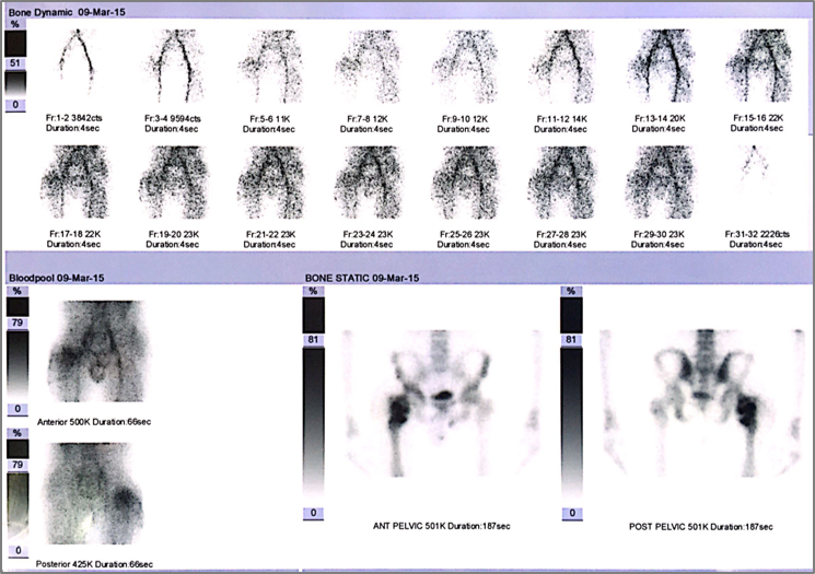
7. A 45-year-old male with history of recent renal transplantation (24 hours ago) and complaint of anuria after transplantation. The images are shown below:
What is the most likely diagnosis? A. Accelerated rejection. B. Acute Tubular Necrosis. C. Urinoma. D. Renal vascular thrombosis.
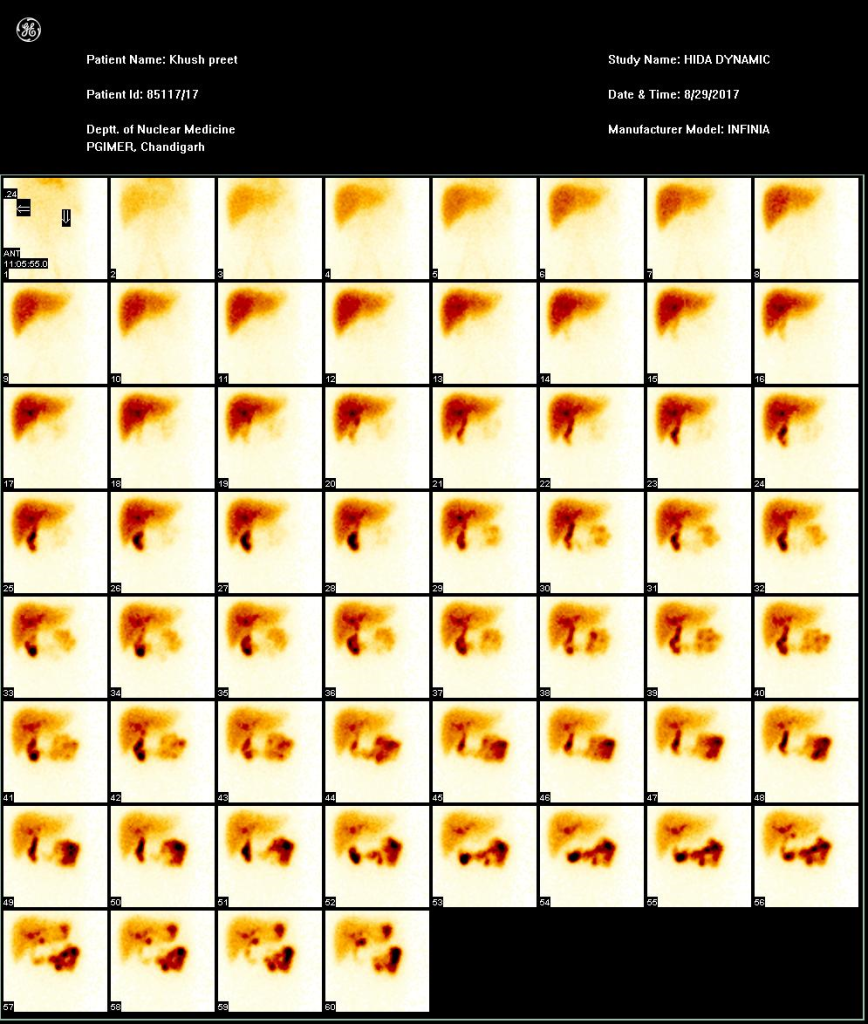
8. What is the abnormal finding indicated by arrow in this HIDA scintigraphy?
A. Duodeno-gastric reflux B. Liver hemangioma C. Bile leak D. Renal activity
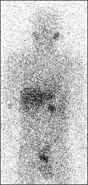
9. A 72-yearold male patient complained fever, diarrhea, and abdominal pain. He underwent Tc-99m labeled leukocyte scan. What is the most likely diagnosis?
A. Infected ulcerative colitis B. Retroperitoneal abscess C. Pyogenic myoscitis D. Lymphoma
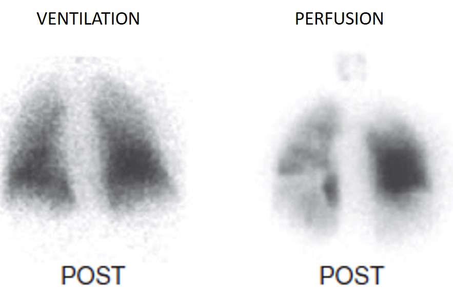
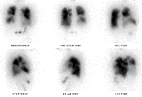
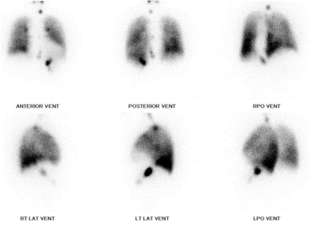
10. A 55-year-old male with acute dyspnoea and cough, normal chest X-ray and the below ventilation/perfusion scan.
What is the probability for pulmonary embolism according to modified PIOPED II criteria? A. High B. Intermediate C. Low D. Very low
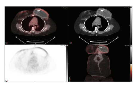
11. Regarding the following images, what is the significance of the FDG uptake seen?
A. Recurrence in area of prosthesis B. Inflammation around prosthesis C. Brown fat uptake D. Cutaneous lymphoma
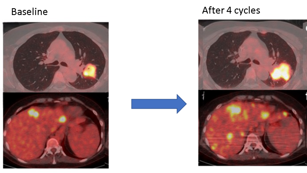
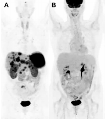
12. A 45-year-old woman with metastatic lung carcinoma treated with nivolumab. The following images show F-18 FDG PET/CT images from baseline and after 4 cycles of therapy. Based on the findings of the images, which of the following statement is true?
A. Scan findings are suggestive of partial response B. Scan findings are suggestive of disease progression C. Scan findings are suggestive of unconfirmed progression D. Scan findings are suggestive of stable disease.
13. A 49-year-old lady presented with fever of unknown origin. She underwent FDG PET-CT for further evaluation (image). Based on the PET-CT findings, what is the most likely cause of her fever?
A. Esophagitis B. Vasculitis C. Pyelonephritis D. Esophageal carcinoma
14. What is the most likely diagnosis regarding the following scan images?
A. Well differentiated Neuroendocrine tumour Grade 1 B. Well differentiated Neuroendocrine tumour Grade 2 C. Well differentiated Neuroendocrine tumour Grade 3 D. Neuroendocrine Carcinoma
15. A 63-year-old male, a known case of papillary carcinoma thyroid. Post thyroidectomy, post 150 mCi of radio iodine (I-131) in 2008, post neck dissection for recurrent disease, post 150 mCi of radio iodine (I-131) in 2014. Patient was lost to follow up and recently complained of mild dyspnoea and there was increase in his serum thyroglobulin levels on evaluation. His whole-body radioiodine scan and whole-body F-18 FDG PET-CT scan images are given below.
What is best line of treatment for this patient ? A. High dose (150-200 mCi) radio-iodine (iodine-131) therapy in view of pulmonary metastasis B. Lenvatinib / other tyrosine kinase inhibitors in view of radio iodine refractory status C. Chemotherapy D. Best supportive care
16. The landmark VISION trial demonstrated the safety and efficacy of which therapeutic radiopharmaceutical?
A. Lu-177 PSMA-617 B. Ac-225 PSMA-617 C. Lu- 177 DOTA-TATE D. Ra-223 Chloride
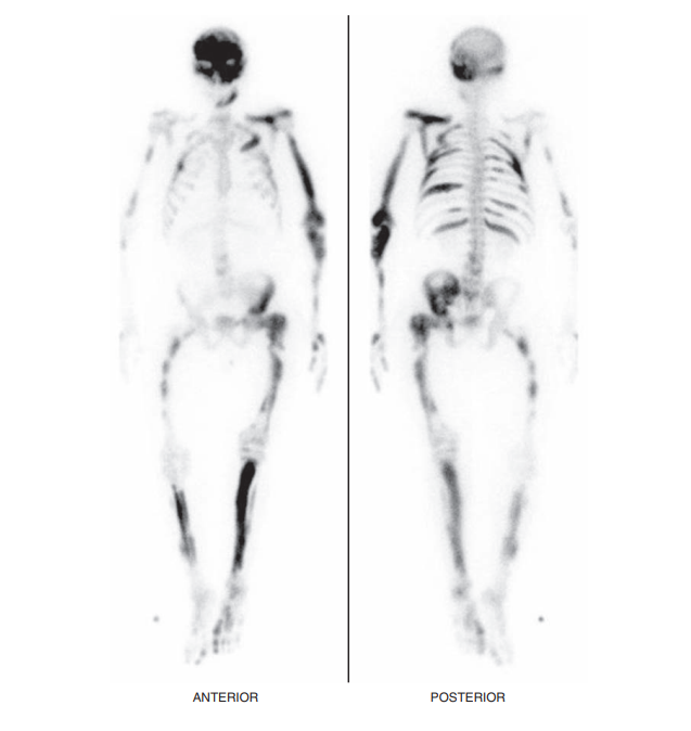
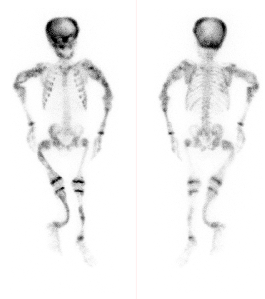
17. Considering the following image, what is the most likely diagnosis?
A. Polyostotic fibrous dysplasia B. Osseous metastases C. Osteomyelitis D. Osteochondromas
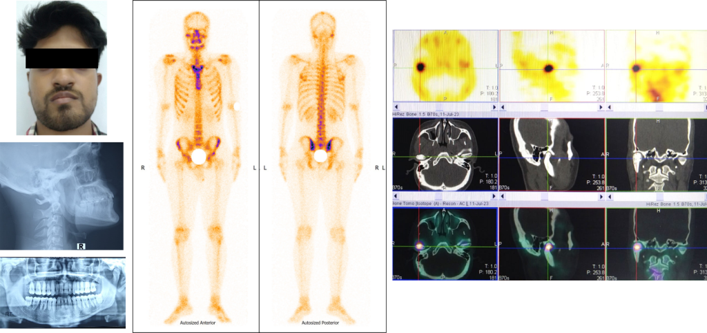
18. A 22-year-male was presented with facial asymmetry for few months. Recently he had noticed for pain at right temporomandibular region and few episodes of lock jaw. Plain and OPG X-ray was done and then he was advised for Tc-99m MDP bone scan (Clinical picture, Plain and OPG X-ray, Whole body bone scan and SPECT-CT scan images are shown below):
What is the most likely diagnosis?
A. Osteochondroma at condyle of right mandible B. Unilateral condylar hyperplasia of right mandible C. Osteoarthritis of right temporomandibular joint D. Osteoarthrosis of right temporomandibular region
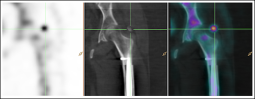
19. A 35-year-old woman is in a motor vehicle accident and suffers multiple fractures, including traumatic cervical spine injury. Three months following her accident she begins to experience lower limb spasms. What is the most likely diagnosis based on her delayed bone scan SPECT/CT?
A. Displaced bone fragment after femoral fracture B. Greater trochanter enthesopathy C. Heterotopic ossification D. Trochanteric bursitis
20. A 10-year-old girl with pain in her right lower extremity on F-18 FDG PET showed a primary tumor (likely osteosarcoma) in the proximal right tibia and soft tissue. There was increased FDG uptake in ipsilateral inguinal femoral lymphnode and was considered reactive. The nuclear physician is interested to compare F-18 FDG uptake with another PET tracer. Which one of the following PET tracers is currently being used in sarcomas other than the F-18 FDG PET?
A. Tb-161 DOTATOC B. C-11 choline C. Ga-68 FAPI D. F-18 Flouroestradiol
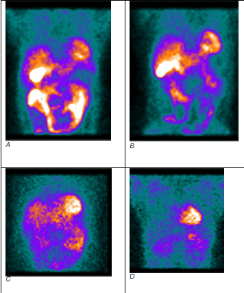
21. A 54-year-old male patient has benign sporadic adrenal pheochromocytoma. Nuclear scans are shown in the figure. Which radiopharmaceuticals were used to image scans labeled as A, B, C, and D in the figure?
A. F-18 FDG, Ga-68 DOTA SSA, F-18 FDOPA and I-123 MIBG B. F-18 FDG, Ga-68 DOTA SSA, F-18 FLT and I-123 octreotide C. Ga-68 DOTA SSA, F-18 FDOPA, I-123 MIBG and F-18 FDG D. Ga-68 DOTA SSA, F-18 FLT, I-123 MIBG and F-18 FDG
22. A 40-year-old male presented with right limb lower swelling for one year. A lymphoscintigraphy was done following injection of 99mTc sulphur colloid (images shown below):
Which of the following statements is true?
A. Scan findings are suggestive of complete lymphatic obstruction in both lower limbs. B. Scan findings are suggestive of complete lymphatic obstruction in right lower limb and partial lymphatic obstruction in left lower limb. C. Scan findings are suggestive of partial lymphatic obstruction in both lower limbs. D. Scan findings are suggestive of partial lymphatic obstruction in right lower limb.
23. A three- and half-year male child underwent Tc-99m DMSA (a) and Tc-99m EC scan (b) within a week. Biochemical analysis revealed normal serum creatinine. What is the prime imaging differential?
A. Tracer extravasation in DMSA study B. Proximal renal tubular acidosis C. Chronic renal failure D. Pyelonephritis
24. Identify the PET radiopharmaceutical in the following Maximal Intensity Projection image?
A. F-18 Fluciclovine B. Ga-68 Exendin C. Ga-68 RM2 D. Ga-68 RGD
25. A 35-year-old female with a history of gradually increasing lethargy and multiple episodes of dizziness over the past 6 years underwent a biochemical workup that showed elevated levels of plasma insulin and C-peptide. Patient underwent Ga-68 DOTA-Exendin PET/CT imaging. Based on the findings of the scan, what is the most likely explanation for the observed appearance?
A. Nesidioblastosis B. Insulinoma C. VIPoma D. Gastrinoma
Sample Questions from the 2018 Exam
1. From the images of lung perfusion and ventilation studies, which of the following is the most likely probability for pulmonary embolism in this case?
a.High
b.Intermediate
c. Low
d.Very low
2. Assessment of the presynaptic dopamine transporter with SPECT is helpful for the differential diagnosis between:
A. Essential tremor and parkinsonian syndromes
B. Parkinson’s disease and multiple system atrophy
C. DOPA-responsive dystonia and essential tremor
D. Familial Parkinson’s disease and vascular Parkinsonism
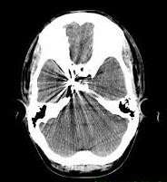
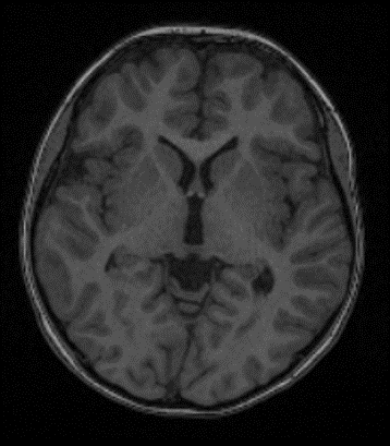
3. Choose the correct sentence:
A. Cerebellum is shown in this slice
B. There is midline shift.
C. This patient has hydrocephalus.
C. Third ventricle is shown in this slice.
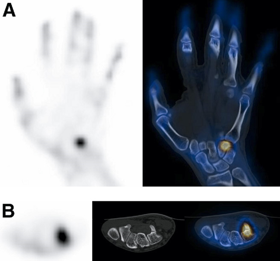
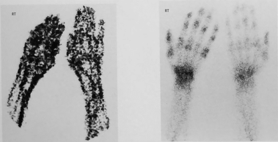
4. A 25 yrs old man complains of wrist pain and referred for bone scan. The images is shown below. What is the most probable diagnosis? (ANATOMY)
A. AVN of scaphoid
B .Fracture of hamate
C. Keinbock Disease
D. Fracture of triquetrum
5. The full width at half maximum (FWHM) of a photo-peak is a measure of:
A. PHA window setting
B. Camera sensitivity
C. Field of view
D. Detector energy resolution
6. Arrange the following PET tracers in the increasing order of their half-lives:
A. O-15, C-11, F-18, N-13
B. C-11, O-15, N-13, F-18
C. O-15, C-11, N-13, F-18
D. O-15, N-13, C-11, F-18
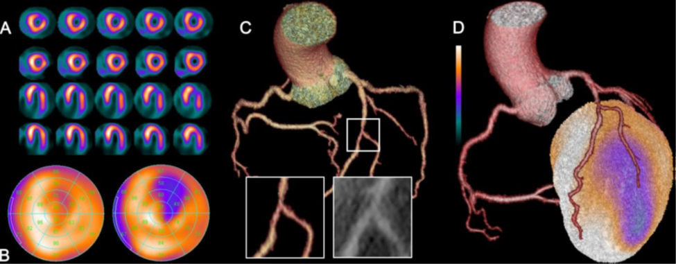
7. The picture shows:
A. Myocardial ischemia in anteroseptal wall with corresponding critical stenosis of first diagonal artery.
B. Myocardial ischemia in the anterolateral wall with non-significant stenosis due to calcification in D1 bifurcation.
C. Myocardial ischemia in the anterolateral wall due to intermediate stenosis by non-calcified plaques in artery at the level of the second diagonal branch bifurcation.
D. Myocardial infarction with peri-infarction ischemia in the anterior wall due to occlusion of D2 bifurcation
8. A 30-year-old male with hypertension underwent a nuclear medicine test. What is the test and diagnosis?
A. 99mTc-DMSA Renal cortical scintigraphy-renovascular hypertension
B. 131I-MIBG scintigraphy-pheochromocytoma
C. 99mTc-MIBI parathyroid scintigraphy-renal calculi
D. 201Tl scintigraphy-left renal cell carcinoma
9. A case of mediastinal lymphoma status post chemoradiotherapy, what is the most likely diagnosis in this fused PET/CT image.
A. Radiation induced pneumonitis
B. Lymphangitis carcinamatosis
C. Fungal chest infection
D. Drug induced pneumonitis
10. Cellular hypoxia is assessed by:
A. 18F-FDG
B. 11C-Acetate
C. 15O-Water
D. 18F-Fluromisonidazole
11. A 60-year-old male patient had received bisphosphonate therapy against bone metastasis of prostate cancer. The patient underwent FDG PET-CT. Which is the diagnosis for the jaw finding?
A. Metastasis
B. Primary cancer
C. EB virus infection
D. Osteonecrosis
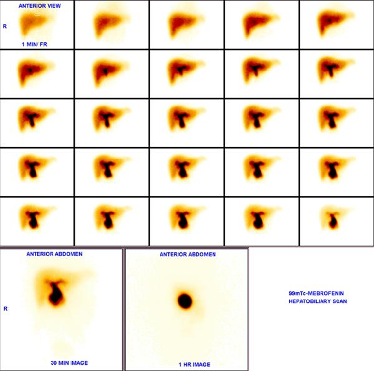
12. Diagnosis of the following scan is:
A. Acute cholecystitis
B. Chronic Cholecystitis
C. Hepatobiliary leak
D. Choledochal cyst
13. An 8-year-old boy presented with features suggesting precocious puberty, brown patches on face & chest and abnormality of upper and lower limbs. His physician suspected genetic syndrome and referred him for 99mTc MDP bone scan (as shown below). These findings can be best explained as:
A. Paget’s disease
B. Osteomalacia
C. Extensive metastases
D. Fibrous dysplasia
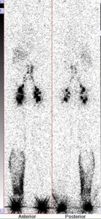
14. A 45-year-old man with right arm and hand pain was referred for 3 phase bone scan, which is shown below. What is the most probable diagnosis?
A. Diffuse Cellulitis
B. Degenerative arthritis
C. Rhematoid arthritis
D. Complex Regional Pain Syndrome
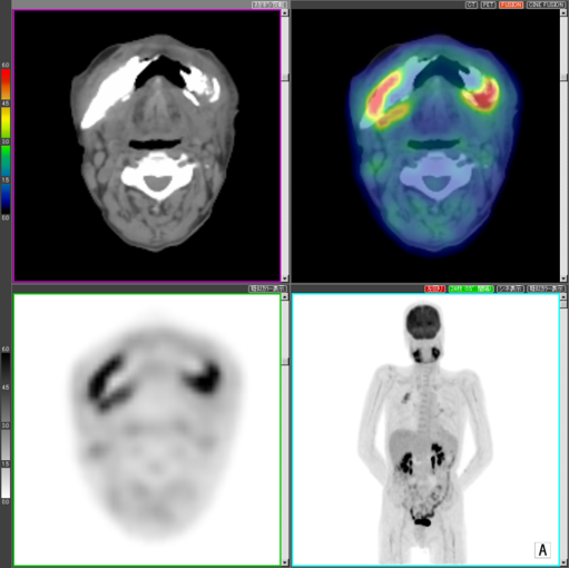
15. A 60-year-old woman with locally advanced lung adenocarcinoma presents for staging FDG PET/CT scan. What is the best explanation for the FDG uptake indicated by the arrow?
A. Synchronous primary carcinoma in the head and neck
B. Physiologic FDG uptake
C. Compensatory vocal cord uptake
D. Vocalisation during uptake phase of the scan
16. A 60-year-old male with a history of right hip pain underwent a 3-phase bone scan (shown below). All of the choices listed below can be considered in the differential diagnosis except:
A. Osteomyelitis
B. Osteosarcoma
C. Arthritis
D. Healing fracture
17. Given the myocardial perfusion scan (MPS) of a 50-year-old female (see below) with a TID of 1.1 which of the following statements is true for the image below:
A. Normal MPI
B. Balanced ischemia
C. Transient ischemic dilatation
D. Suspected triple-vessel disease
18. In renal transplant evaluation, Acute Tubular Necrosis typically is associated with:
A. Poor perfusion, slow but good uptake
B. Poor perfusion, poor uptake
C. Good perfusion, slow but good uptake
D. Good perfusion, poor uptake
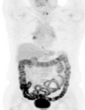
19. What is the most likely cause of the bowel uptake in the following PET scan?
A. Insulin administration
B. Metformin
C. Colon cancer
D. Bowel ischemia
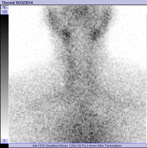
20. A 30-year-old female presents with a goiter and palpitations of two weeks’ duration. Serum FT4 is 50 (normal value of 12 to 22), TSH is 0.1 (normal value of 0.3 to 3.5). Radioactive iodine (I-131) uptake is 1.3% at 24 hours. Thyroid scan is shown below. The most probable diagnosis is: